
Gall
Galls (from the Latin galla, 'oak-apple') or cecidia (from the Greek kēkidion, anything gushing out) are a kind of swelling growth on the external tissues of plants. Plant galls are abnormal outgrowths[1] of plant tissues, similar to benign tumors or warts in animals. They can be caused by various parasites, from viruses, fungi and bacteria, to other plants, insects and mites. Plant galls are often highly organized structures so that the cause of the gall can often be determined without the actual agent being identified. This applies particularly to insect and mite plant galls. The study of plant galls is known as cecidology.
Anatomy[edit]
Shape and size[edit]
Galls develop on various plant organs, providing nutrition and shelter to inducing insects. Galls display vast variation in morphology, size, and wall composition. The size of insect galls can range significantly, from approximately two inches in diameter to less than one-sixteenth of an inch. Some galls are so small that they are merely slightly thickened patches on leaves [2]. Their shape can range from spherical to bursiform, bullet-shaped, flower-shaped, cylindrical, or diamond-like. Factors influencing gall morphology include plant species, tissue type, gall-inducing agent, and environmental conditions[3][4][5][6][7]. They typically exhibit symmetrical forms, although their end shapes vary due to differences in the physical actions and chemical stimuli of different insects. Around 90% of galls occur on the leaves of dicotyledons[8]. Galls can develop on various parts of the host plant, such as roots, leaf bases, branches, or leaflets. Internally, galls also exhibit diverse structures. Some are simple, comprising only outgrown and curved leaf tissues, while others feature complex, hierarchical arrangements with multiple chambers containing different types of tissues, including collenchyma, parenchyma, physalides-parenchyma, and a nutritive cellular layer[9][10][11].
Structure[edit]
In a general gall wasp gall, the outermost layer is the epidermis followed by outer cortex and then inner cortex. In some galls these two cortex layers are separated by a lignified layer. The innermost part of a gall is the larval chamber. The nutritive layer situated between the larval chamber and the inner cortex. There is a nutritional gradient (high to low) from inside to outside of the gall while defense gradient to the opposite direction[12][13].
Morphogenesis[edit]
Gall morphogenesis involves the regulation of the organ on which the gall occurs while maintaining differentiation freedom. Gall development begins from a single or group of metaplasied cells and progresses through promoter-mediated cell expansion, cell multiplication, programmed differentiation, and control of symmetry[8].
Plant response involves the establishment of metaplasied cells and localized metabolic changes to repair the wound and neutralize stress. Osmotic stress leads to the development of metaplasied cells, characterized by increased quantities of osmotically active material. The rejection response by the plant triggers the synthesis of defense compounds and enzymes[14][15].
Differentiation[edit]
- Development of novel cell types: Galls exhibit unique cell types such as abnormally thick-walled dead cells (e.g., xylary elements and sclereids) and thin-walled living cells. These cells differentiate in specific patterns, contributing to the structure of the gall[16][17][18].
- Nutritive tissue: Most galls contain specialized nutritive tissue that provides nutrition to the inducing arthropod and sometimes to their progeny. The structure of this tissue varies depending on the insect species inducing the gall and their feeding behaviors. Nutritive tissue differentiation is influenced by the length and nature of the insect's mouthparts[19].
- Characteristics of nutritive cells: Nutritive cells exhibit dynamic features such as enriched cytoplasm, fragmented vacuoles, hypertrophied nucleus and nucleolus, and abundant cell organelles. They contain elevated levels of carbohydrates, lipids, soluble sugars, and proteins, along with intense phosphatase activity[20].
- Changes in nutritive tissue: The activity of the nutritive tissue is maintained as long as the inhabiting larva continues to feed. However, when feeding ceases, the dynamic profile of the tissue gradually diminishes, and it is eventually replaced by inactive parenchyma. Removal or death of the larva leads to rapid changes in the distribution of carbohydrates and lipids within the tissue[21].
- Accumulation of phenolic substances: Cells lining the larval chamber in mature-old galls accumulate phenolic substances, indicating changes in gall tissue composition over time[22].
- Mineral content: Gall tissues contain elevated levels of various minerals, which may play a role in gall development and function[23][4].
Types[edit]
There are two primary categories of galls: closed and open[2]. Insects such as wasps, moths, and flies, possessing chewing mouthparts during their adult or larval stages, typically inhabit completely enclosed galls. Upon reaching maturity, the adult exits either by chewing its way out or utilizing an opening created by the larval stage. Conversely, insects with sucking mouthparts rely on partially open galls or those that naturally open to facilitate emergence. An example of the latter type is the aphid, which forms marble-sized galls on the leaf stems of cottonwood trees. While these galls have thin walls, they harbor entire colonies of aphids within. When the time is right, a slit appears on one side of the gall, allowing the aphids to escape as the slit's lips unfold[3][18].
Physiology[edit]
Insects induce the formation of galls on plants from which they receive various services, such as a source of nutrition and a place to lay eggs, develop, and be provided protection from the environment and enemies. The gall producers are specific to specific plants, thus inducing galls with unique appearances (balls, knobs, lumps, warts, etc.) and a range of colors (red, green, yellow, and black). Different taxonomic groups of gall inducers vary in the complexity and diversity of gall formation and organization, with insect induced galls generally being more complex and diverse[24]. Additionally, gall frequency varies based on factors such as weather, plant susceptibility, and pest populations.
Gall Formation[edit]
There are four stages of gall development: initiation, growth and differentiation, maturation, and dehiscence. Gall tissues are nutritive and present high concentrations of lipids, proteins, nitrogen, and other nutrients.
The formation of galls which is induction begins with insect saliva on plants. Insect saliva contains various chemicals, induces shock and osmotic changes in the host plant cell[8]. The severity of insect feeding injures the plant varies depending on the insect. The osmotic changes that occur as a result are characterized by increased quantities of osmotically active material and induce the cell metaplasia and gall formation.
Gall growth occurs gradually over time, with the length, breadth, and height of the galls increasing proportionally. The growth rate is maximal during the insect's early developmental stages and slows as it approaches adulthood. Hormones like auxins play a crucial role in gall growth. The presence of stress and insect secretions stimulates the synthesis of growth-promoting substances, possibly involving a combination of different growth promoters like auxins and kinins. Gall growth involves both cell enlargement and division, but the specific factors triggering cell enlargement remain unclear[25][26].
The earliest impact from the insect leads to metaplasia in the affected cells, where they undergo changes in structure and function. When the chemical shock is of high intensity, metaplasia does not occur. Instead, the plant cells local to the shock die, thereby rejecting the insect and defending the plant tissue. Enzymes like invertases are involved in gall growth, with greater activity correlating with stronger gall development. Gall-inducing insect performance is influenced by plant vigor and module size, with larger, fast-growing plant modules resulting in larger galls. Conversely, galls are easily induced on smaller plant modules[4][8][13][23].
Genetics[edit]
Galls are unique growths on plants, and how the plant's genetic instructions could produce these structures in response to external factors is still a fresh field of science. Genetic mechanisms of gall formation is a unique interplay between the parasite and the host plant in shaping the developmental trajectory of the gall organ.
The 'zigzag' model introduced by Jones & Dangl (2006)[27] demonstrates the molecular interactions underlying gall induction. This model, refined over time and subject to ongoing enhancements, illustrates the intricate dynamics between antagonistic molecular players. Pattern-triggered immunity (PTI), constitutes the initial defense layer of plant cells, activated upon detection of "danger signals." These signals, termed damage-associated-molecular-patterns (DAMPs) if originating from the plant or microbe/pathogen-associated-molecular-patterns (MAMPs, PAMPs, or HAMPs)[28] if from the parasite, engage pattern-recognition receptors (PRRs) triggering signaling cascades. PRRs, classified as receptor-like kinases (RLKs), mediate intercellular communication by bridging external stimuli with intracellular defense mechanisms. Antagonists, employing effector-triggered susceptibility (ETS) manipulate host-cell functions through effector molecules encoded by effector genes, aiming primarily at suppressing plant defenses. Notably, some effectors exploit plant traits, known as "plant susceptibility traits," diverting the plant's resources in favor of the parasite. Effectoromics, involving high-throughput expression screens, aids in identifying effector candidates crucial for colonization. Conversely, Effector-Triggered Immunity (ETI) responsible for plant's counterattack, leveraging effectors as "danger signals" to render the parasite avirulent. During ETI, nucleotide-binding domain leucine-rich repeat (NLR)-containing receptors detect perturbations induced by effectors, leading to downstream signaling events that promote defense responses. However, parasites can counteract ETI by modifying ETS, undermining the efficacy of resistance genes deployed in agriculture. The evolutionary arms race between plants and parasites, underscored by the expansion of gene families involved in biotic interactions, shapes their genomic landscape, influencing their adaptive strategies and diversification[29][30].
Crown galls formed under the influence of the bacterium Agrobacterium tumefaciens exhibit several distinctive characteristics when compared to other types of galls. This bacterium transfers genetic material known as T-DNA into the plant cells, where it becomes integrated into the chromosomes. The T-DNA contains genes that encode for production of auxin, cytokinin and opines. As a result, the infected plant cells undergo rapid multiplication, essentially transforming into "bacterial factories" that produce more bacterial bodies[2].
Certain bacteria, like Rhodococcus fascians, induce the formation of leafy galls on plants, affecting their growth. These galls act as permanent sinks, diverting nutrients away from other parts of the plant and causing growth suppression elsewhere. The bacteria possess virulence genes that control their ability to colonize plants and produce cytokinins, which influence plant growth. While parasitic gall-inducers are typically harmful to plants, researchers are exploring ways to harness their growth-promoting abilities for agricultural benefit. Some derivatives of R. fascians are being investigated for their potential to promote balanced plant growth, and scientists are also studying plant interactions with these bacteria to discover traits that could enhance crop yields.
Most of the transcriptomic studies on plant galls used entire gall samples resulting both gall and non-gall cells leading to thousands of gene expressions during gall development[31][32]. Recent studies on gall induced by gall wasps (Hymenoptera: Cynipidae)[33] Dryocosmus quercuspalustris on northern red oak (Quercus rubra L.) leaves demonstrate the complexity of genetic mechanisms underlying galls by quantifying the tissue-specific gene expression[34]. There are substantial differences in gene expression between inner and outer gall tissues compared to adjacent leaf tissues. Notably, approximately 28% of oak genes display differential expression in the gall compared to leaves, indicating significant transcriptional changes associated with gall development[34]. According to the transcriptome analysis, while the outer gall transcriptome resembles that of twigs, leaf buds, and reproductive structures, the inner gall transcriptome is distinct from normal oak tissues, underscoring the complexity of gall formation[12]. Furthermore, there is an upregulation of genes related to sugar and amino acid metabolism in both outer and inner gall tissues, suggesting a role in transporting plant metabolites to support the nutritional needs of the developing gall wasp larva. The defense-related genes are found to be suppressed in inner gall tissues as a strategy to accommodate the feeding activity of the parasite[35].
Taxonomic range[edit]
Plant galls are caused by a wide range of organisms, including animals such as insects, mites, and nematodes; fungi; bacteria; viruses; and other plants.
Insects[edit]
Insect galls are the highly distinctive plant structures formed by some herbivorous insects as their own microhabitats. They are plant tissue which is controlled by the insect. Galls act as both the habitat and food source for the maker of the gall. The interior of a gall can contain edible nutritious starch and other tissues. Some galls act as "physiologic sinks", concentrating resources in the gall from the surrounding plant parts.[26] Galls may also provide the insect with physical protection from predators.[36][25]
Insect galls are usually induced by chemicals injected by the larvae of the insects into the plants and possibly mechanical damage. After the galls are formed, the larvae develop inside until fully grown, when they leave. To form galls, the insects must take advantage of the time when plant cell division occurs quickly: the growing season, usually spring in temperate climates, but which is extended in the tropics.
The meristems, where plant cell division occurs, are the usual sites of galls, though insect galls can be found on other parts of the plant, such as the leaves, stalks, branches, buds, roots, and even flowers and fruits. Gall-inducing insects are usually species-specific and sometimes tissue-specific on the plants they gall.
Gall-inducing insects include gall wasps, gall midges, gall flies, leaf-miner flies, aphids, scale insects, psyllids, thrips, gall moths, and weevils.[37]
Many gall insects remain to be described. Estimates range up to more than 210,000 species, not counting parasitoids of gall-forming insects.[38]
Cynipid wasps[edit]
More than 1400 species of cynipid wasps cause galls. Some 1000 of these are in the tribe Cynipini, their hosts mostly being oak trees and other members of the Fagaceae (the beech tree family).[38] These are often restricted taxonomically to a single host species or a group of related species.
- Cynipid wasp galls
-
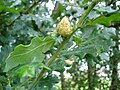 Oak artichoke gall caused by Andricus foecundatrix
 -
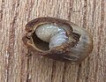 Artichoke gall cut open to reveal wasp larva
 -
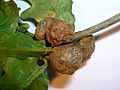
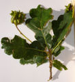 Knopper gall caused by Andricus quercuscalicis
 -
 Oak gall caused by Neuroterus albipes forma laeviusculus
 -
 Marble gall of oak caused by Andricus kollari
 -
 Cherry oak gall caused by Cynips quercusfolii
 -
 Cherry oak gall cut open to reveal wasp larva
 -
 Cherry oak gall wasp adult
 -
 Red-pea gall (Cynips divisa) on pedunculate oak
 -
 Cola-nut galls (Andricus lignicola) on pedunculate oak
 -
 Kokkocynips rileyi oak gall
 -
 Phylloteras poculum oak galls


.jpg)


Non-cynipid wasps[edit]
Some wasps from other groups, such as the Diplolepididae and the Chalcidoidea, also cause plant galls.
- Non-cynipid wasp galls
-

-
 Section through young bedeguar gall showing wasp larvae and cells
 -


Hemipteran bugs[edit]
Among the hemipteran bugs that cause galls are the psyllid bug Pachypsylla celtidisumbilicus, and the woolly aphid Adelges abietis, which parasitises coniferous trees such as the Sitka spruce and the Norway spruce.
- Hemipteran galls
-
 Developing pineapple pseudocone galls on Norway spruce, caused by woolly aphid Adelges abietis
 -
 Pineapple gall cut open to show the woolly aphid larvae inside
 -
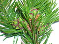
 Pineapple gall on Sitka spruce caused by Adelges abietis
 -
 Pachypsylla celtidisumbilicus hackberry gall


Dipteran flies[edit]
Some dipteran flies such as the cecidomyiid gall midges Dasineura investita and Neolasioptera boehmeriae, and some Agromyzidae leaf-miner flies cause galls.
- Midge galls
-

-
 Nettle gall caused by Dasineura investita (Cecidomyiidae)
 -
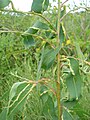
 False nettle stem gall caused by gall midge Neolasioptera boehmeriae (Cecidomyiidae)
 -
 Schizomyia impatientis (Cecidomyiidae) jewelweed flower gall


Mites[edit]
Mites, small arachnids, cause distinctive galls in plants such as the lime tree.
- Mite galls
-
 Lime nail galls caused by the mite Eriophyes tiliae

Nematodes[edit]
Nematodes are microscopic worms that live in soil. Some nematodes (Meloidogyne species or root-knot nematodes) cause galls on the roots of susceptible plants. The galls are often small.[39][40]
- Nematode galls
-
 Juvenile Meloidogyne penetrating a host plant
 -
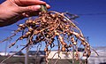 Root-knot galls caused by the nematode Meloidogyne
_penetrates_a_tomato_root_-_USDA-ARS.jpg)

Fungi[edit]
Many rust fungi induce gall formation, including western gall rust, which infects a variety of pine trees and cedar-apple rust. Galls are often seen in Millettia pinnata leaves and fruits. Leaf galls appear like tiny clubs; however, flower galls are globose. Exobasidium often induces spectacular galls on its hosts.
The fungus Ustilago esculenta associated with Zizania latifolia, a wild rice, produces an edible gall highly valued as a food source in the Zhejiang and Jiangsu provinces of China.[41]
- Fungal galls
-

-
 Leaf galls on Rhododendron ferrugineum caused by fungus Exobasidium rhododendri


Bacteria and viruses[edit]
Gall-causing bacteria include Agrobacterium tumefaciens and Pseudomonas savastanoi.
Gall forming virus was found on rice plants in central Thailand in 1979 and named rice gall dwarf. Symptoms consisted of gall formation along leaf blades and sheaths, dark green discoloration, twisted leaf tips, and reduced numbers of tillers. Some plants died in the glasshouse in the later stages of infection. The causal agent was transmitted by the hemipteran bug Nephotettix nigropictus after an incubation of two weeks. Polyhedral particles of 65 nm diameter in the cytoplasm of phloem cells were always associated with the disease. No serologic relationship was found between this virus and that of rice dwarf.
- Microbial pathogen galls
-
 Crown gall on Kalanchoe infected with Agrobacterium tumefaciens
 -
 Citrus vein enation woody gall on Fortunella japonica caused by a pathogen with an aphid vector

_on_Fortunella_japonica.jpg)
Bacteria crown galls
Plants[edit]
The hemiparasitic plant mistletoe forms woody structures sometimes called galls on its hosts.[42] More complex interactions are possible; the parasitic plant Cassytha filiformis sometimes preferentially feeds on galls induced by the cynipid wasp Belonocnema treatae.[43]
Physiology of insect-induced galls[edit]
This section needs additional citations for verification. (May 2024) |
Insects induce the formation of galls on plants from which they receive various services, such as a source of nutrition and a place to lay eggs, develop, and be provided protection from the environment and enemies. The gall producers are specific to specific plants, thus inducing galls with unique appearances (balls, knobs, lumps, warts, etc.) and a range of colors (red, green, yellow, and black). Different taxonomic groups of gall inducers vary in the complexity and diversity of gall formation and organization, with insect induced galls generally being more complex and diverse.[44] Additionally, gall frequency varies based on factors such as weather, plant susceptibility, and pest populations.
There are four stages of gall development: initiation, growth and differentiation, maturation, and dehiscence. Gall tissues are nutritive and present high concentrations of lipids, proteins, nitrogen, and other nutrients. The formation of galls begins with insect saliva on plants inducing a chemical shock.[45] The osmotic changes that occur as a result are characterized by increased quantities of osmotically active material and induce the cell metaplasia and gall formation. When the chemical shock is of high intensity, metaplasia does not occur. Instead, the plant cells local to the shock die, thereby rejecting the insect and defending the plant tissue.
Uses[edit]
Galls are rich in resins and tannic acid and have been used widely in the manufacturing of permanent inks (such as iron gall ink) and astringent ointments, in dyeing, and in leather tanning. The Talmud[46] records using gallnuts as part of the tanning process as well as a dye-base for ink.
Medieval Arabic literature records many uses for the gall, called ˁafṣ in Arabic. The Aleppo gall, found on oak trees in northern Syria, was among the most important exports from Syria during this period, with one merchant recording a shipment of galls from Suwaydiyya near Antioch fetching the high price of 4½ dinars per 100 pounds. The primary use of the galls was as a mordant for black dyes; they were also used to make a high-quality ink. The gall was also used as a medication to treat fever and intestinal ailments.[47]
See also[edit]
- British Plant Gall Society
- Forest pathology
- List of insect galls
- Similar structures:
- Burl
- Witch's broom
References[edit]
- ^ "gall(4)", Merriam-Webster Online Dictionary, accessed November 16, 2007: "an abnormal outgrowth of plant tissue usually due to insect or mite parasites or fungi and sometimes forming an important source of tannin".
- ^ a b c "TPWD: Plant Galls -- Young Naturalist". tpwd.texas.gov. Retrieved 2024-05-09.
- ^ a b Krikorian, A. D. (June 1988). "Plant Galls and Gall Inducers. Jean Meyer , S. Cheskin". The Quarterly Review of Biology. 63 (2): 225–226. doi:10.1086/415876. ISSN 0033-5770.
- ^ a b c Barnes, Jeffrey K. (1993-01-01). "Biology of Insect-Induced Galls". Annals of the Entomological Society of America. 86 (1): 122–123. doi:10.1093/aesa/86.1.122. ISSN 1938-2901.
- ^ Crespi, Bernard; Worobey, Michael (December 1998). "Comparative Analysis of Gall Morphology in Australian Gall Thrips: The Evolution of Extended Phenotypes". Evolution. 52 (6): 1686–1696. doi:10.1111/j.1558-5646.1998.tb02248.x. ISSN 0014-3820.
- ^ Heard, Stephen B.; Buchanan, Corinne K. (October 1998). "Larval Performance and Association Within and Between Two Species of Hackberry Nipple Gall Insects, Pachypsylla spp. (Homoptera: Psyllidae)". The American Midland Naturalist. 140 (2): 351–357. doi:10.1674/0003-0031(1998)140[0351:lpaawa]2.0.co;2. ISSN 0003-0031.
- ^ Florentine, S. K.; Raman, A.; Dhileepan, K. (October 2005). "Effects of Gall Induction by Epiblema Strenuana on Gas Exchange, Nutrients, and Energetics in Parthenium Hysterophorus". Biocontrol. 50 (5): 787–801. doi:10.1007/s10526-004-5525-3. hdl:1959.17/64564. ISSN 1386-6141.
- ^ a b c d Raman, Anantanarayanan (2011-06-01). "Morphogenesis of insect-induced plant galls: facts and questions". Flora - Morphology, Distribution, Functional Ecology of Plants. 206 (6): 517–533. doi:10.1016/j.flora.2010.08.004. ISSN 0367-2530.
- ^ Arduin, M.; Kraus, J.E. (1995-06-25). "Anatomia e Ontogenia de Galhas Foliares de Piptadenia gonoacantha (Fabales, Mimosaceae)". Boletim de Botânica. 14: 109. doi:10.11606/issn.2316-9052.v14i0p109-130. ISSN 2316-9052.
- ^ KRAUS, JANE E.; ARDUIN, MARCOS; VENTURELLI, MARGARIDA (December 2002). "Anatomy and ontogenesis of hymenopteran leaf galls of Struthanthus vulgaris Mart. (Loranthaceae)". Revista Brasileira de Botânica. 25 (4): 449–458. doi:10.1590/s0100-84042002012000009. ISSN 0100-8404.
- ^ Maresquelle, H. J.; Meyer, J. (1965), "Physiologie et morphogenèse des galles d'origine animale (zoocécidies)", Differenzierung und Entwicklung / Differentiation and Development, Berlin, Heidelberg: Springer Berlin Heidelberg, pp. 1927–1976, doi:10.1007/978-3-642-50088-6_49, ISBN 978-3-642-50090-9, retrieved 2024-05-08
- ^ a b Schultz, Jack C.; Stone, Graham N. (June 2022). "A tale of two tissues: Probing gene expression in a complex insect-induced gall". Molecular Ecology. 31 (11): 3031–3034. doi:10.1111/mec.16482. ISSN 0962-1083. PMC 9321127. PMID 35466464.
- ^ a b Williams, Michele A J, ed. (1994-10-06). Plant Galls: Organisms, Interactions, Populations. Oxford University PressOxford. doi:10.1093/oso/9780198577690.001.0001. ISBN 978-0-19-857769-0.
- ^ Carmen, Cogălniceanu Gina, "Electrical Control of Plant Morphogenesis", Plant Tissue Culture Engineering, Berlin/Heidelberg: Springer-Verlag, pp. 397–415, doi:10.1007/1-4020-3694-9_21 (inactive 2024-05-09), ISBN 1-4020-3594-2, retrieved 2024-05-09
{{citation}}: CS1 maint: DOI inactive as of May 2024 (link) - ^ Sinnott, Edmund W. (1960). Plant morphogenesis. New York: McGraw-Hill. doi:10.5962/bhl.title.4649.
- ^ Gasson, P (September 2000). "Fink S. 1999.Pathological and regenerative plant anatomy. Encyclopedia of plant anatomy XIV. 1095 pp. Berlin, Stuttgart: Gebrüder Borntraeger". Annals of Botany. 86 (3): 707–708. doi:10.1006/anbo.2000.1242. ISSN 0305-7364.
- ^ Maresquelle, H. J.; Meyer, J. (1965), "Physiologie et morphogenèse des galles d'origine animale (zoocécidies)", Differenzierung und Entwicklung / Differentiation and Development, Berlin, Heidelberg: Springer Berlin Heidelberg, pp. 1927–1976, doi:10.1007/978-3-642-50088-6_49, ISBN 978-3-642-50090-9, retrieved 2024-05-09
- ^ a b ROHFRITSCH, O.; SHORTHOUSE, J.D. (1982), "Insect Galls", Molecular Biology of Plant Tumors, Elsevier, pp. 131–152, doi:10.1016/b978-0-12-394380-4.50011-6, ISBN 978-0-12-394380-4, retrieved 2024-05-09
- ^ Bronner, R.; Westphal, E.; Dreger, F. (February 1989). "Chitosan, a component of the compatible interaction between Solanum dulcamara L. and the gall mite Eriophyes cladophthirus Nal". Physiological and Molecular Plant Pathology. 34 (2): 117–130. doi:10.1016/0885-5765(89)90020-9. ISSN 0885-5765.
- ^ Gasson, P (September 2000). "Fink S. 1999.Pathological and regenerative plant anatomy. Encyclopedia of plant anatomy XIV. 1095 pp. Berlin, Stuttgart: Gebrüder Borntraeger". Annals of Botany. 86 (3): 707–708. doi:10.1006/anbo.2000.1242. ISSN 0305-7364.
- ^ Schwartz, W. (1966). "M. S. Mani, Ecology of Plant Galls (Monogr. Biol. Vol. XII). 434 u. XII S., 164 Abb., 9 Taf. The Hague 1964: Dr. W. Junk Publishers. 40.-hfl". Zeitschrift für allgemeine Mikrobiologie. 6 (1): 91. doi:10.1002/jobm.3630060116 (inactive 2024-05-09). ISSN 0044-2208.
{{cite journal}}: CS1 maint: DOI inactive as of May 2024 (link) - ^ Jensen, P. Boysen (January 1948). "Formation of Galls by Mikiola fagi". Physiologia Plantarum. 1 (1): 95–108. doi:10.1111/j.1399-3054.1948.tb07113.x. ISSN 0031-9317.
- ^ a b Mapes, Carol C. (2005), "Gall Formation", Encyclopedia of Entomology, Dordrecht: Springer Netherlands, pp. 942–944, doi:10.1007/0-306-48380-7_1732, ISBN 978-0-306-48380-6, retrieved 2024-05-09
- ^ Gatjens-Boniche, Omar (2019-12-01). "The mechanism of plant gall induction by insects: revealing clues, facts, and consequences in a cross-kingdom complex interaction". Revista de Biología Tropical. 67 (6): 1359–1382. doi:10.15517/rbt.v67i6.33984. ISSN 2215-2075.
- ^ a b Stone, G. N.; Schonrogge, K. (2003) "The adaptive significance of insect gall morphology", Trends in Ecology & Evolution 18(10): 512–522. doi:10.1016/S0169-5347(03)00247-7.
- ^ a b Larson, K. C.; Whitham, T. G. (1991). "Manipulation of food resources by a gall-forming aphid: the physiology of sink-source interactions", Oecologia 88(1): 15–21. doi:10.1007/BF00328398.
- ^ Jones, Jonathan D. G.; Dangl, Jeffery L. (November 2006). "The plant immune system". Nature. 444 (7117): 323–329. doi:10.1038/nature05286. ISSN 0028-0836. PMID 17108957.
- ^ Zipfel, Cyril; Felix, Georg (August 2005). "Plants and animals: a different taste for microbes?". Current Opinion in Plant Biology. 8 (4): 353–360. doi:10.1016/j.pbi.2005.05.004. ISSN 1369-5266. PMID 15922649.
- ^ Voinnet, Olivier (2005-02-10). "Induction and suppression of RNA silencing: insights from viral infections". Nature Reviews Genetics. 6 (3): 206–220. doi:10.1038/nrg1555. ISSN 1471-0056. PMID 15703763.
- ^ Kessler, André; Baldwin, Ian T. (June 2002). "P<scp>LANT</scp> R<scp>ESPONSES TO</scp> I<scp>NSECT</scp> H<scp>ERBIVORY</scp>: The Emerging Molecular Analysis". Annual Review of Plant Biology. 53 (1): 299–328. doi:10.1146/annurev.arplant.53.100301.135207. ISSN 1543-5008. PMID 12221978.
- ^ Betancourt, Eunice Kariñho; Soto, Paulina Hernández; Cortés, Nancy Calderón; Anaya, Martha Rendón; Estrella, Alfredo Herrera; Oyama, Ken (2020), "Ecological Genomics of Plant-Insect Interactions: The Case of Wasp-Induced Galls", Evolutionary Ecology of Plant-Herbivore Interaction, Cham: Springer International Publishing, pp. 315–341, doi:10.1007/978-3-030-46012-9_17, ISBN 978-3-030-46011-2, retrieved 2024-05-09
- ^ Hearn, Jack; Blaxter, Mark; Schönrogge, Karsten; Nieves-Aldrey, José-Luis; Pujade-Villar, Juli; Huguet, Elisabeth; Drezen, Jean-Michel; Shorthouse, Joseph D.; Stone, Graham N. (2019-11-04). "Genomic dissection of an extended phenotype: Oak galling by a cynipid gall wasp". PLOS Genetics. 15 (11): e1008398. doi:10.1371/journal.pgen.1008398. ISSN 1553-7404. PMC 6855507. PMID 31682601.
- ^ Sunnucks, Paul; Stonet, G N; Schonrogget, K; Csokat, G (1994-10-06), "The biogeography and population genetics of the invading gall wasp Andricus quercuscalicis (Hymenoptera: Cynipidae)", Plant Galls, Oxford University PressOxford, pp. 351–368, doi:10.1093/oso/9780198577690.003.0021, ISBN 978-0-19-857769-0, retrieved 2024-05-09
- ^ a b Martinson, Ellen O.; Werren, John H.; Egan, Scott P. (June 2022). "Tissue-specific gene expression shows a cynipid wasp repurposes oak host gene networks to create a complex and novel parasite-specific organ". Molecular Ecology. 31 (11): 3228–3240. doi:10.1111/mec.16159. ISSN 0962-1083. PMID 34510608.
- ^ Weis, Arthur E.; Abrahamson, Warren G. (1986). "Evolution of Host-Plant Manipulation by Gall Makers: Ecological and Genetic Factors in the Solidago-eurosta System". The American Naturalist. 127 (5): 681–695. doi:10.1086/284513. ISSN 0003-0147. JSTOR 2461318.
- ^ Weis, A. E.; Kapelinski, A. (1994). "Variable selection on Eurosta's gall size. II. A path analysis of the ecological factors behind selection", Evolution 48(3): 734–745. doi:10.1111/j.1558-5646.1994.tb01357.x.
- ^ Volovnik, S. V. (2010). "Weevils Lixinae (Coleoptera, Curculionidae) as Gall Formers", Entomological Review, 90(5): 585–590. doi:10.1134/S0013873810050052.
- ^ a b Brandão-Dias, Pedro F. P.; Zhang, Yuanmeng Miles; Pirro, Stacy; Vinson, Camila C.; et al. (2022). "Describing biodiversity in the genomics era: A new species of Nearctic Cynipidae gall wasp and its genome". Systematic Entomology. 47 (1). Wiley: 94–112. doi:10.1111/syen.12521. ISSN 0307-6970.
- ^ Krusberg, L. R. (1963). "Host Response to Nematode Infection". Annual Review of Phytopathology. 1 (1). Annual Reviews: 219–240. doi:10.1146/annurev.py.01.090163.001251. ISSN 0066-4286.
- ^ Williamson, V. M.; Hussey, R. S. (1996). "Nematode pathogenesis and resistance in plants". The Plant Cell. 8 (10). Oxford University Press: 1735–1745. doi:10.1105/tpc.8.10.1735. ISSN 1040-4651. PMC 161311. PMID 8914324.
- ^ Terrell, E. E.; Batra, L. R. "Zizania latifolia and Ustilago esculenta, a grass-fungus association", Economic Botany 36(3): 274–285. doi:10.1007/BF02858549.
- ^ "Modes of union and interaction between parasite and host in the Loranthaceae. III. Further observations on Viscum and Korthalsella". Proceedings of the Royal Society of London. Series B - Biological Sciences. 148 (931). The Royal Society: 188–206. 18 February 1958. doi:10.1098/rspb.1958.0013. ISSN 2053-9193.
- ^ Egan, Scott P.; Zhang, Linyi; Comerford, Mattheau; Hood, Glen R. (August 2018). "Botanical parasitism of an insect by a parasitic plant". Current Biology. 28 (16): R863–R864. doi:10.1016/j.cub.2018.06.024. PMID 30130501.
- ^ Gatjens-Boniche, Omar (2019-12-01). "The mechanism of plant gall induction by insects: revealing clues, facts, and consequences in a cross-kingdom complex interaction". Revista de Biología Tropical. 67 (6): 1359–1382. doi:10.15517/rbt.v67i6.33984. ISSN 2215-2075.
- ^ Raman, Anantanarayanan (2011-06-01). "Morphogenesis of insect-induced plant galls: facts and questions". Flora - Morphology, Distribution, Functional Ecology of Plants. 206 (6): 517–533. doi:10.1016/j.flora.2010.08.004. ISSN 0367-2530.
- ^ Bavli, tractate Gittin:19a
- ^ Goitein, Shelomo Dov; Sanders, Paula (1967). A Mediterranean Society: Daily life. University of California Press. p. 405. ISBN 0520048695. Retrieved 22 June 2020.
Further reading[edit]
- Blanche, Rosalind (2012). Life in a Gall: The Biology and Ecology of Insects that Live in Plant Galls. Collingwood, Vic.: CSIRO Publishing. ISBN 978-0643106437.
- Redfern, Margaret (2011). Plant Galls. London: Collins. ISBN 978-0002201445.
- Russo, Ron (2007). Field Guide to Plant Galls of California and Other Western States. Berkeley, California: University of California Press. ISBN 978-0520248854.
External links[edit]
- British Plant Gall Society
- A Field Guide to Plant Galls of the North East U.S.
- To Be or Not To Be a Gall: The Story of Strange Growths on Plants Archived 2014-03-21 at the Wayback Machine
- Insect Galls. Brandeis University
- "Common oak galls". University of Kentucky Entomology. Archived from the original on 2006-09-13. Retrieved 2006-09-11.
| International | |
|---|---|
| National | |