
User:Colin/Rotavirus
| Rotavirus | |
|---|---|

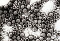
| |
| Computer reconstruction of a transmission electron micrograph of a rotavirus particle. | |
| Virus classification | |
| Group: | Group III (dsRNA)
|
| Family: | |
| Genus: | Rotavirus
|
| Species: | A,B,C,D,E,F, and G
|
Rotavirus is a genus of double-stranded RNA virus in the taxonomic family Reoviridae. It is the leading cause of severe diarrhoea among infants and young children; by the age of five, nearly every child in the world has been infected with rotavirus at least once.[1] Rotavirus was discovered in 1973,[2] and is now known to account for up to 50% of infants and children being hospitalised with severe diarrhoea.[3]
There are seven species of rotavirus, referred to as A, B, C, D, E, F, and G. Rotavirus A, the most prevalent, causes more than 90% of infections in humans. Within Rotavirus A there are different strains called serotypes.[4] Rotavirus also infects animals and is an important pathogen of livestock.[5]
Classification[edit]
There are seven species of rotavirus, referred to as A, B, C, D, E, F, and G. Humans are primarily infected by species A, B and C, most commonly by species A. All seven species cause disease in animals.[4]
- Rotavirus species
-
 Rotavirus A in the faeces of an infected child
 -
 Rotavirus B particles
 -
 A higher magnification of rotavirus C particles reacting with antibodies – the blue bar = 100nm


Within rotavirus A there are different strains, called serotypes.[6] Two genes determine these serotypes, and a strain of rotavirus A is classified both by its G and P type. Serotyping of rotavirus A is thus complex; each virus has a P-type and a G-type. P-type is indicated by a number for the P-serotype and by a number in square brackets for the coresponding P-genotype. G-serotypes and are similarly numbered but the G-genotype number is the same as the G-serotype. For example, the rotavirus strain Wa is defined as P1A[8]G1. This dual classification system is similar to that used for influenza virus.[7] Because the genes that determine G-types and P-types can be passed on separately to offspring various, combinations occur in any one strain.[8]
Structure[edit]
The genome of rotavirus consists of eleven unique double helix molecules of RNA which are 18,555 nucleoside base-pairs in total. Each helix, or segment, is a gene, numbered 1 to 11 by decreasing size. Each gene codes for one protein, except genes 9 and 11, which each code for two proteins.[9] The RNA is surrounded by a three-layered icosahedral protein capsid. Viral particles are up to 76.5 nm in diameter[10][11] and are not enveloped.
Proteins[edit]


There are six viral proteins (VPs), that form the virus particle (virion). These structural proteins are called VP1, VP2, VP3, VP4, VP6 and VP7. In addition to the VPs, there are six non-structural proteins (NSPs), that are only produced in cells infected by rotavirus. These are called NSP1, NSP2, NSP3, NSP4, NSP5 and NSP6.
At least six of the twelve proteins encoded by the rotavirus genome bind RNA.[12] The role of these proteins play in rotavirus replication is not entirely understood, their functions are thought to be related to RNA synthesis and packaging in the virion, mRNA transport to the site of genome replication, and mRNA translation and regulation of gene expression.[13]
Structural proteins[edit]
VP1 is located in the core of the virus particle and is an RNA polymerase enzyme.[14] In an infected cell this enzyme produces mRNA transcripts for the synthesis of viral proteins and produces copies of the rotavirus genome RNA segments for newly produced virus particles.
VP2 forms the core layer of the virion and binds the RNA genome.[15]
VP3 is part of the inner core of the virion and is an enzyme called guanylyl transferase. This is a capping enzyme that catalyses the formation of the 5' cap in the post-transcriptional modification of mRNA.[16] The cap stabilises viral mRNA by protecting it from nucleic acid degrading enzymes called nucleases, and is required for mRNA export to the cytoplasm.
VP4 is on the surface of the virion that protrudes as a spike.[17] VP4 has many functions. It binds to molecules on the surface of cells called receptors and drives the entry of the virus into the cell.[18] VP4 has to be modified by a protease enzyme, (found in the gut), into VP5* and VP8* before the virus is infectious.[19] It determines how virulent the virus is and it determines the P-type of the virus.[20]
VP6 forms the bulk of the capsid. It is highly antigenic and can be used to identify rotavirus species.[21] This protein is used in laboratory tests for rotavirus A infections.[22]
VP7 is a glycoprotein that forms the outer surface of the virion. Apart from its structural functions, it determines the G-type of the strain and, along with VP4, is important in immunity to infection.[10]
Non-structural viral proteins[edit]
NSP1, the product of gene 5, is a nonstructural RNA-binding protein.[23]
NSP2 is an RNA-binding protein that accumulates in cytoplasmic inclusions (viroplasms) and is required for genome replication.[24][25]
NSP3 is bound to viral mRNAs in infected cells and it is responsible for the shutdown of cellular protein synthesis.[26]
NSP4 is a viral enterotoxin to induce diarrhoea and was the first viral enterotoxin discovered.[27]
NSP5 is encoded by genome segment 11 of rotavirus A and in virus-infected cells NSP5 accumulates in the viroplasm.[28]
Gene 11 encodes NSP6, from an out of phase open reading frame.[29] NSP6 is a nucleic acid binding protein.[30]
| RNA Segment (Gene) | Size (base pairs) | Protein | Molecular weight kDa | Location | Function |
|---|---|---|---|---|---|
| 1 | 3302 | VP1 | 125 | At the vertices of the core | RNA-dependent RNA polymerase |
| 2 | 2690 | VP2 | 102 | Forms inner shell of the core | Stimulates viral RNA replicase |
| 3 | 2591 | VP3 | 88 | At the vertices of the core | Guanylyl transferase mRNA capping enzyme |
| 4 | 2362 | VP4 | 87 | Surface spike | Cell attachment, virulence, |
| 5 | 1611 | NSP1 | 59 | Non-structural | Not essential to virus growth |
| 6 | 1356 | VP6 | 45 | Inner Capsid | Structural and species-specific antigen |
| 7 | 1104 | NSP3 | 37 | Non-structural | Enhances viral mRNA translation |
| 8 | 1059 | NSP2 | 35 | Non-structural | NTPase involved in RNA packaging |
| 9 | 1062 | VP71 VP72 | 38 and 34 | Surface | Structural and neutralisation antigen |
| 10 | 751 | NSP4 | 20 | Non-structural | Enterotoxin |
| 11 | 667 | NSP5 NSP6 | 22 | Non-structural | ssRNA and dsRNA binding modulator of NSP2 |
This table is based on the simian rotavirus strain SA11.[31][32] RNA-protein coding assignments differ in some strains.
Replication[edit]

Rotavirus infects enterocytes of the villi of the small intestine, leading to structural and functional changes of the epithelium.[33] Their triple protein coats make them resistant to the acidic pH of the stomach, and the digestive enzymes in the gut.
They enter cells by receptor mediated endocytosis and form a vesicle known as an endosome. Proteins in the third layer (VP7 and the VP4 spike) disrupt the membrane of the endosome, creating a difference in the calcium concentration. This causes the breakdown of VP7 trimers into single protein subunits, leaving the VP2 and VP6 protein coats around the viral dsRNA, forming a double-layered particle (DLP).
The eleven dsRNA strands remain within the protection of the two protein shells and the viral RNA-dependent RNA polymerase creates mRNA transcripts of the double-stranded viral genome. By remaining in the core the viral RNA evades innate host immune responses called RNA interference that are triggered by the presence of double-stranded RNA.
During the infection, rotavirus produces mRNA for both protein biosynthesis and gene replication. Most of the rotavirus proteins accumulate in viroplasm, where the RNA is replicated and the DLPs are assembled. Viroplasm is formed as early as two hours after virus infection around the cell nucleus and, are viral factories and are thought to be made by two viral non-structural proteins, NSP5 and NSP2. Inhibition of NSP5 by RNA interference results in a sharp decrease in rotavirus replication. The DLPs migrate to the endoplasmic reticulum where they obtain their third, outer layer (formed by VP7 and VP4). The progeny viruses are released from the cell by lysis.[34][35]
History[edit]

In 1943, Jacob Light and Horace Hodes proved that an infectious agent causing scours in cattle was a virus.[36] Three decades later, preserved samples of that virus were shown to be rotavirus.[37] In the intervening years, a virus in mice[38] was shown to be related to the virus causing scours.[39] In 1973, related viruses were described by Ruth Bishop in children with gastroenteritis, in Australia.[2][40]
In 1974, Thomas Henry Flewett suggested the name rotavirus after observing that, viewed through an electron microscope, a rotavirus particle looks like a wheel (rota in Latin).[41][42] This name was later adopted by the International Committee on Taxonomy of Viruses. In 1976, related viruses were described in several other species of animals,[39] these viruses causing acute gastroenteritis were recognised as a collective pathogen affecting humans and animals world-wide.[41] Rotavirus serotypes were first described in 1980.[43] In 1981, rotavirus from humans was first grown in cell cultures derived from monkey kidneys, by adding trypsin to the culture medium.[44] The ability to grow rotavirus in culture accelerated the pace of research, and by the mid-1980s the first candidate vaccines were being evaluated.[45]
See also[edit]
References[edit]
- ^ Velázquez FR, Matson DO, Calva JJ, Guerrero L, Morrow AL, Carter-Campbell S, Glass RI, Estes MK, Pickering LK, Ruiz-Palacios GM (1996). "Rotavirus infections in infants as protection against subsequent infections". N. Engl. J. Med. 335 (14): 1022–8. PMID 8793926.
{{cite journal}}: CS1 maint: multiple names: authors list (link) - ^ a b
Bishop RF, Davidson GP, Holmes IH, Ruck BJ (1973). "Virus particles in epithelial cells of duodenal mucosa from children with acute non-bacterial gastroenteritis". Lancet. 2 (7841): 1281–3. PMID 4127639.
{{cite journal}}: CS1 maint: multiple names: authors list (link) - ^ Rheingans RD, Heylen J, Giaquinto C (2006). "Economics of rotavirus gastroenteritis and vaccination in Europe: what makes sense?". Pediatr. Infect. Dis. J. 25 (1 Suppl): S48–55. PMID 16397429.
{{cite journal}}: CS1 maint: multiple names: authors list (link) - ^ a b Beards GM, Brown DW (1988). "The antigenic diversity of rotaviruses: significance to epidemiology and vaccine strategies". Eur. J. Epidemiol. 4 (1): 1–11. PMID 2833405.
- ^ Holland RE (1990). "Some infectious causes of diarrhea in young farm animals" (PDF). Clin. Microbiol. Rev. 3 (4): 345–75. PMID 2224836.
- ^ Santos N, Hoshino Y (2005). "Global distribution of rotavirus serotypes/genotypes and its implication for the development and implementation of an effective rotavirus vaccine". Rev. Med. Virol. 15 (1): 29–56. doi:10.1002/rmv.448. PMID 15484186.
- ^ Desselberger U, Iturriza-Gómara M, Gray JJ (2001). "Rotavirus epidemiology and surveillance". Novartis Found. Symp. 238: 125–47, discussion 147–52. PMID 11444024.
{{cite journal}}: CS1 maint: multiple names: authors list (link) - ^ Desselberger U, Wolleswinkel-van den Bosch J, Mrukowicz J, Rodrigo C, Giaquinto C, Vesikari T (2006). "Rotavirus types in Europe and their significance for vaccination". Pediatr. Infect. Dis. J. 25 (1 Suppl): S30–41. PMID 16397427.
{{cite journal}}: CS1 maint: multiple names: authors list (link) - ^ Chan WK, Penaranda ME, Crawford SE, Estes MK (1986). "Two glycoproteins are produced from the rotavirus neutralization gene". Virology. 151 (2): 243–52. PMID 3010552.
{{cite journal}}: CS1 maint: multiple names: authors list (link) - ^ a b Pesavento JB, Crawford SE, Estes MK, Prasad BV (2006). "Rotavirus proteins: structure and assembly". Curr. Top. Microbiol. Immunol. 309: 189–219. PMID 16913048.
{{cite journal}}: CS1 maint: multiple names: authors list (link) - ^ Prasad BV, Chiu W (1994). "Structure of rotavirus". Curr. Top. Microbiol. Immunol. 185: 9–29. PMID 8050286.
- ^ Patton JT (1995). "Structure and function of the rotavirus RNA-binding proteins" (PDF). J. Gen. Virol. 76 ( Pt 11): 2633–44. PMID 7595370.
- ^ Patton JT (2001). "Rotavirus RNA replication and gene expression". Novartis Found. Symp. 238: 64–77, discussion 77–81. PMID 11444036.
- ^ Vásquez-del Carpió R, Morales JL, Barro M, Ricardo A, Spencer E (2006). "Bioinformatic prediction of polymerase elements in the rotavirus VP1 protein". Biol. Res. 39 (4): 649–59. PMID 17657346.
{{cite journal}}: CS1 maint: multiple names: authors list (link) - ^ Arnoldi F, Campagna M, Eichwald C, Desselberger U, Burrone OR (2007). "Interaction of rotavirus polymerase VP1 with nonstructural protein NSP5 is stronger than that with NSP2". J. Virol. 81 (5): 2128–37. doi:10.1128/JVI.01494-06. PMID 17182692.
{{cite journal}}: CS1 maint: multiple names: authors list (link) - ^ Fresco LD, Buratowski S (1994). "Active site of the mRNA-capping enzyme guanylyltransferase from Saccharomyces cerevisiae: similarity to the nucleotidyl attachment motif of DNA and RNA ligases" (PDF). Proc. Natl. Acad. Sci. U.S.A. 91 (14): 6624–8. PMID 8022828.
- ^ Gardet A, Breton M, Fontanges P, Trugnan G, Chwetzoff S (2006). "Rotavirus spike protein VP4 binds to and remodels actin bundles of the epithelial brush border into actin bodies". J. Virol. 80 (8): 3947–56. doi:10.1128/JVI.80.8.3947-3956.2006. PMID 16571811.
{{cite journal}}: CS1 maint: multiple names: authors list (link) - ^ Arias CF, Isa P, Guerrero CA, Méndez E, Zárate S, López T, Espinosa R, Romero P, López S (2002). "Molecular biology of rotavirus cell entry". Arch. Med. Res. 33 (4): 356–61. PMID 12234525.
{{cite journal}}: CS1 maint: multiple names: authors list (link) - ^ Konno T, Suzuki H, Kitaoka S, Sato T, Fukuhara N, Yoshie O, Fukudome K, Numazaki Y (1993). "Proteolytic enhancement of human rotavirus infectivity". Clin. Infect. Dis. 16 Suppl 2: S92–7. PMID 8384014.
{{cite journal}}: CS1 maint: multiple names: authors list (link) - ^ Hoshino Y, Jones RW, Kapikian AZ (2002). "Characterization of neutralization specificities of outer capsid spike protein VP4 of selected murine, lapine, and human rotavirus strains". Virology. 299 (1): 64–71. PMID 12167342.
{{cite journal}}: CS1 maint: multiple names: authors list (link) - ^ Bishop RF (1996). "Natural history of human rotavirus infection". Arch. Virol. Suppl. 12: 119–28. PMID 9015109.
- ^ Beards GM, Campbell AD, Cottrell NR, Peiris JS, Rees N, Sanders RC, Shirley JA, Wood HC, Flewett TH (1984). "Enzyme-linked immunosorbent assays based on polyclonal and monoclonal antibodies for rotavirus detection" (PDF). J. Clin. Microbiol. 19 (2): 248–54. PMID 6321549.
{{cite journal}}: CS1 maint: multiple names: authors list (link) - ^ Hua J, Mansell EA, Patton JT (1993). "Comparative analysis of the rotavirus NS53 gene: conservation of basic and cysteine-rich regions in the protein and possible stem-loop structures in the RNA". Virology. 196 (1): 372–8. doi:10.1006/viro.1993.1492. PMID 8395125.
{{cite journal}}: CS1 maint: multiple names: authors list (link) - ^ Kattoura MD, Chen X, Patton JT (1994). "The rotavirus RNA-binding protein NS35 (NSP2) forms 10S multimers and interacts with the viral RNA polymerase". Virology. 202 (2): 803–13. doi:10.1006/viro.1994.1402. PMID 8030243.
{{cite journal}}: CS1 maint: multiple names: authors list (link) - ^ Taraporewala ZF, Patton JT (2004). "Nonstructural proteins involved in genome packaging and replication of rotaviruses and other members of the Reoviridae". Virus Res. 101 (1): 57–66. doi:10.1016/j.virusres.2003.12.006. PMID 15010217.
- ^ Poncet D, Aponte C, Cohen J (1993). "Rotavirus protein NSP3 (NS34) is bound to the 3' end consensus sequence of viral mRNAs in infected cells" (PDF). J. Virol. 67 (6): 3159–65. PMID 8388495.
{{cite journal}}: CS1 maint: multiple names: authors list (link) - ^ Dong Y, Zeng CQ, Ball JM, Estes MK, Morris AP (1997). "The rotavirus enterotoxin NSP4 mobilizes intracellular calcium in human intestinal cells by stimulating phospholipase C-mediated inositol 1,4,5-trisphosphate production". Proc. Natl. Acad. Sci. U.S.A. 94 (8): 3960–5. PMID 9108087.
{{cite journal}}: CS1 maint: multiple names: authors list (link) - ^ Afrikanova I, Miozzo MC, Giambiagi S, Burrone O (1996). "Phosphorylation generates different forms of rotavirus NSP5". J. Gen. Virol. 77 ( Pt 9): 2059–65. PMID 8811003.
{{cite journal}}: CS1 maint: multiple names: authors list (link) - ^ Mohan KV, Atreya CD (2001). "Nucleotide sequence analysis of rotavirus gene 11 from two tissue culture-adapted ATCC strains, RRV and Wa". Virus Genes. 23 (3): 321–9. PMID 11778700.
- ^ Rainsford EW, McCrae MA (2007). "Characterization of the NSP6 protein product of rotavirus gene 11". doi:10.1016/j.virusres.2007.06.011. PMID 17658646.
{{cite journal}}: Cite journal requires|journal=(help) - ^ Desselberger U. Rotavirus: basic facts. In Rotaviruses Methods and Protocols. Ed. Gray, J. and Desselberger U. Humana Press, 2000, pp. 1–8. ISBN 0-89603-736-3
- ^ Patton JT. Rotavirus RNA replication and gene expression. In Novartis Foundation. Gastroenteritis Viruses, Humana Press, 2001, pp. 64–81. ISBN 0-471-49663-4
- ^ Greenberg HB, Clark HF, Offit PA (1994). "Rotavirus pathology and pathophysiology". Curr. Top. Microbiol. Immunol. 185: 255–83. PMID 8050281.
{{cite journal}}: CS1 maint: multiple names: authors list (link) - ^ Jayaram H, Estes MK, Prasad BV (2004). "Emerging themes in rotavirus cell entry, genome organization, transcription and replication". Virus Res. 101 (1): 67–81. doi:10.1016/j.virusres.2003.12.007. PMID 15010218.
{{cite journal}}: CS1 maint: multiple names: authors list (link) - ^ Patton JT, Vasquez-Del Carpio R, Spencer E (2004). "Replication and transcription of the rotavirus genome". Curr. Pharm. Des. 10 (30): 3769–77. PMID 15579070.
{{cite journal}}: CS1 maint: multiple names: authors list (link) - ^ Light JS, Hodes HL (1943). "Studies on Epidemic Diarrhea of the New-born: Isolation of a Filtrable Agent Causing Diarrhea in Calves". Am J Public Health Nations Health. 33 (12): 1451–1454. PMID 18015921.
- ^ Mebus CA, Wyatt RG, Sharpee RL; et al. (1976). "Diarrhea in gnotobiotic calves caused by the reovirus-like agent of human infantile gastroenteritis" (PDF). Infect. Immun. 14 (2): 471–4. PMID 184047.
{{cite journal}}: Explicit use of et al. in:|author=(help)CS1 maint: multiple names: authors list (link) - ^
Rubenstein D, Milne RG, Buckland R, Tyrrell DA (1971). "The growth of the virus of epidemic diarrhoea of infant mice (EDIM) in organ cultures of intestinal epithelium". British journal of experimental pathology. 52 (4): 442–45. PMID 4998842.
{{cite journal}}: CS1 maint: multiple names: authors list (link) - ^ a b
Woode GN, Bridger JC, Jones JM, Flewett TH, Davies HA, Davis HA, White GB (1976). "Morphological and antigenic relationships between viruses (rotaviruses) from acute gastroenteritis in children, calves, piglets, mice, and foals" (PDF). Infect. Immun. 14 (3): 804–10. PMID 965097.
{{cite journal}}: CS1 maint: multiple names: authors list (link) Cite error: The named reference "pmid965097" was defined multiple times with different content (see the help page). - ^
Bishop RF, Davidson GP, Holmes IH, Ruck BJ (1973). "Letter: Evidence for viral gastroenteritis". N. Engl. J. Med. 289 (20): 1096–7. PMID 4742237.
{{cite journal}}: CS1 maint: multiple names: authors list (link) - ^ a b Flewett TH, Woode GN (1978). "The rotaviruses". Arch. Virol. 57 (1): 1–23. PMID 77663.
- ^ Flewett TH, Bryden AS, Davies H, Woode GN, Bridger JC, Derrick JM (1974). "Relation between viruses from acute gastroenteritis of children and newborn calves". Lancet. 2 (7872): 61–3. PMID 4137164.
{{cite journal}}: CS1 maint: multiple names: authors list (link) - ^
Beards GM, Pilfold JN, Thouless ME, Flewett TH (1980). "Rotavirus serotypes by serum neutralisation". J. Med. Virol. 5 (3): 231–7. PMID 6262451.
{{cite journal}}: CS1 maint: multiple names: authors list (link) - ^
Urasawa T, Urasawa S, Taniguchi K (1981). "Sequential passages of human rotavirus in MA-104 cells". Microbiol. Immunol. 25 (10): 1025–35. PMID 6273696.
{{cite journal}}: CS1 maint: multiple names: authors list (link) - ^
Vesikari T, Isolauri E, Delem A; et al. (1985). "Clinical efficacy of the RIT 4237 live attenuated bovine rotavirus vaccine in infants vaccinated before a rotavirus epidemic". J. Pediatr. 107 (2): 189–94. PMID 3894608.
{{cite journal}}: Explicit use of et al. in:|author=(help)CS1 maint: multiple names: authors list (link)