
File:Kinetoplast of Trypanosoma brucei.tif

No higher resolution available.
Kinetoplast_of_Trypanosoma_brucei.tif (758 × 369 pixels, file size: 455 KB, MIME type: image/tiff)
| This is a file from the Wikimedia Commons. Information from its description page there is shown below. Commons is a freely licensed media file repository. You can help. |
Summary
| Description |
English: Trypanosoma brucei, Electron micrograph of centrin-depleted cells. (a) A typical control cell in this particular section shows single flagellum, pair of basal bodies, kinetoplast and nucleus. (b) TbCen2-depleted cells with multi basal bodies and the kinetoplast with enlarged size compared to the control in ‘a’. Scale bars, 500 nm. F, flagellum; B, basal body; K, kinetoplast; N, nucleus; P, flagellar pocket. |
| Date | |
| Source | Fig. 3 of: Role of Centrins 2 and 3 in Organelle Segregation and Cytokinesis in Trypanosoma brucei. In: PLOS ONE; doi:10.1371/journal.pone.0045288 |
| Author | Angamuthu Selvapandiyan, Praveen Kumar, Jeffrey L. Salisbury, Ching C. Wang, Hira L. Nakhasi |
Licensing
This file is licensed under the Creative Commons Attribution 2.5 Generic license.
- You are free:
- to share – to copy, distribute and transmit the work
- to remix – to adapt the work
- Under the following conditions:
- attribution – You must give appropriate credit, provide a link to the license, and indicate if changes were made. You may do so in any reasonable manner, but not in any way that suggests the licensor endorses you or your use.
File history
Click on a date/time to view the file as it appeared at that time.
| Date/Time | Thumbnail | Dimensions | User | Comment | |
|---|---|---|---|---|---|
| current | 03:34, 22 January 2015 | 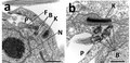 | 758 × 369 (455 KB) | Chhandama | User created page with UploadWizard |
File usage
The following pages on the English Wikipedia use this file (pages on other projects are not listed):
Global file usage
The following other wikis use this file:
- Usage on ar.wikipedia.org
- Usage on bg.wikipedia.org
- Usage on bs.wikipedia.org
- Usage on de.wikipedia.org
- Usage on it.wikipedia.org
- Usage on ja.wikipedia.org
- Usage on nl.wikipedia.org
- Usage on ru.wikipedia.org