
Posterior cord
| Posterior cord | |
|---|---|
 Plan of brachial plexus. (Posterior cord is shaded gray.) | |
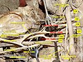
 The right brachial plexus with its short branches, viewed from in front. The Sternomastoid and Trapezius muscles have been completely removed, the Omohyoid and Subclavius have been partially removed; a piece has been sawed out of the clavicle; the Pectoralis muscles have been incised and reflected. | |
| Details | |
| From | brachial plexus - posterior divisions of the three trunks |
| To | subscapular, up. and low. thoracodorsal axillary radial |
| Innervates | none |
| Identifiers | |
| Latin | fasciculus posterior plexus brachialis |
| TA98 | A14.2.03.023 |
| TA2 | 6416 |
| FMA | 45237 |
| Anatomical terms of neuroanatomy | |
The posterior cord is a part of the brachial plexus. It consists of contributions from all of the roots of the brachial plexus.[1]
The posterior cord gives rise to the following nerves:[2]
| Name | Roots | Supplies |
| upper subscapular nerve | C5-C6 | subscapularis muscle of the rotator cuff |
| lower subscapular nerve | C5-C6 | subscapularis muscle, teres major muscle |
| thoracodorsal nerve | C6-C8 | latissimus dorsi muscle |
| axillary nerve | C5-C6 | sensation to the shoulder and motor to the deltoid muscle, the teres minor and the triceps brachii (long head) muscle |
| radial nerve | C5-C8, T1 | triceps brachii muscle, the brachioradialis muscle, the extensor muscles of the fingers and wrist (extensor carpi radialis muscle), supinator, and the extensor and abductor muscles of the thumb |
Additional images[edit]
-
 Brachial plexus
 -
 Posterior cord
 -
 Posterior cord
 -
 Brachial plexus. Deep dissection.
 -
 Brachial plexus. Deep dissection. Anterolateral view


References[edit]
MBBS resources http://mbbsbasic.googlepages.com/
External links[edit]
- Atlas image: hand_plexus at the University of Michigan Health System - "Axilla, dissection, anterior view"
This neuroscience article is a stub. You can help Wikipedia by expanding it. |