
Lesser omentum
| Lesser omentum | |
|---|---|
 | |
 | |
| Details | |
| Identifiers | |
| Latin | omentum minus |
| TA98 | A10.1.02.101 |
| TA2 | 3750 |
| FMA | 9715 |
| Anatomical terminology | |
The lesser omentum (small omentum or gastrohepatic omentum) is the double layer of peritoneum that extends from the liver to the lesser curvature of the stomach, and to the first part of the duodenum. The lesser omentum is usually divided into these two connecting parts: the hepatogastric ligament, and the hepatoduodenal ligament.[1]
Structure[edit]


The lesser omentum is extremely thin, and is continuous with the two layers of peritoneum which cover respectively the antero-superior and postero-inferior surfaces of the stomach and first part of the duodenum.
When these two layers reach the lesser curvature of the stomach and the upper border of the duodenum, they join and ascend as a double fold to the porta hepatis.
To the left of the porta, the fold is attached to the bottom of the fossa for the ductus venosus, along which it is carried to the diaphragm, where the two layers separate to embrace the end of the esophagus.
At the right border of the lesser omentum, the two layers are continuous, and form a free margin which constitutes the anterior boundary of the omental foramen.
Divisions[edit]
Anatomically, the lesser omentum is divided into ligaments, each starting with the prefix "hepato" to indicate that it connects to the liver at one end.
Most sources divide it into two parts:[1]
- hepatogastric ligament: the portion connecting to the lesser curvature of the stomach
- hepatoduodenal ligament: the portion connecting to the duodenum
In some cases, the following ligaments are considered part of the lesser omentum:
- hepatophrenic ligament: the portion connecting to the thoracic diaphragm[2]
- hepatoesophageal ligament: the portion connecting to the esophagus[3]
- hepatocolic ligament: the portion connecting to the colon
Contents[edit]
Between the two layers of the lesser omentum, close to the right free margin, are the hepatic artery proper, the common bile duct, the portal vein, lymphatics, and the hepatic plexus of nerves—all these structures being enclosed in a fibrous capsule (Glisson's capsule).
Between the layers of the lesser omentum, where they are attached to the stomach, run the right and left gastric arteries, as well as the gastric veins.
Additional images[edit]
-
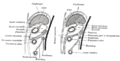 Diagrams to illustrate the development of the greater omentum and transverse mesocolon.
 -
 Horizontal disposition of the peritoneum in the upper part of the abdomen.


See also[edit]
- Omental bursa (Lesser sac)
- Greater sac
- Omental foramen (Epiploic foramen, Foramen of Winslow)
- Greater omentum – Fat sheath under abdominal wall
- Peritoneum – Serous membrane that forms lining of abdominal cavity or coelom
References[edit]
![]() This article incorporates text in the public domain from page 1156 of the 20th edition of Gray's Anatomy (1918)
This article incorporates text in the public domain from page 1156 of the 20th edition of Gray's Anatomy (1918)
- ^ a b abdominalcavity at The Anatomy Lesson by Wesley Norman (Georgetown University) (peritoneumonsagcut, xsectthrulesseromentum)
- ^ Anatomy photo:38:st-0304 at the SUNY Downstate Medical Center - "Stomach, Spleen and Liver: Ligaments"
- ^ Anatomy photo:37:05-0103 at the SUNY Downstate Medical Center - "Abdominal Cavity: The Lesser Omentum"
External links[edit]
- Weiglein, Andreas H. (1996). "Variations and topography of the arteries in the lesser omentum in humans". Clinical Anatomy. 9 (3): 143–150. doi:10.1002/(SICI)1098-2353(1996)9:3<143::AID-CA1>3.0.CO;2-H. PMID 8740472.
- Dux, K. (1990). "Anatomy of the greater and lesser omentum in the mouse with some physiological implications". The Omentum. pp. 19–43. doi:10.1007/978-1-4612-3436-4_3. ISBN 978-1-4612-8011-8.
- Nayak, Satheesha B (2009). "Abnormal peritoneal fold connecting the greater omentum with the liver, gallbladder, right kidney and lesser omentum" (PDF). Bratislavske Lekarske Listy. 110 (11): 736–737. PMID 20120448.
- Anatomy figure: 37:04-01 at Human Anatomy Online, SUNY Downstate Medical Center - "The stomach and lesser omentum."
- Anatomy photo:37:05-0100 at the SUNY Downstate Medical Center - "Abdominal Cavity: The Lesser Omentum"
- Anatomy photo:38:03-0102 at the SUNY Downstate Medical Center - "Stomach, Spleen and Liver: Contents of the Hepatoduodenal Ligament"
- Anatomy image:7823 at the SUNY Downstate Medical Center
- Anatomy image:8147 at the SUNY Downstate Medical Center
- Peritoneal Cavity Development - Page 6 of 14 anatomy module at med.umich.edu