
File:Ebstein4.jpg
Size of this preview: 800 × 507 pixels. Other resolutions: 320 × 203 pixels | 640 × 406 pixels | 847 × 537 pixels.
Original file (847 × 537 pixels, file size: 68 KB, MIME type: image/jpeg)
| This is a file from the Wikimedia Commons. Information from its description page there is shown below. Commons is a freely licensed media file repository. You can help. |
Summary
| Description |
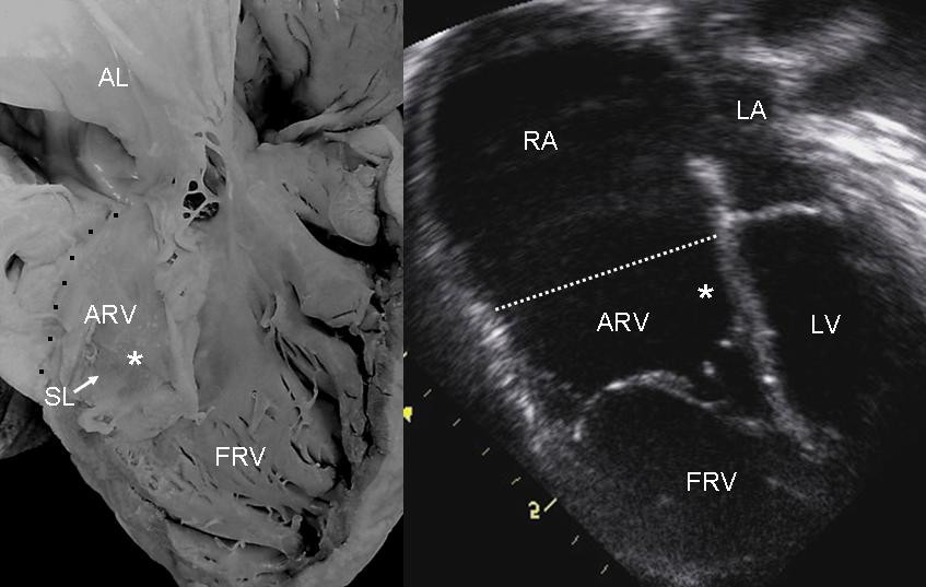
English: Internal view of the right ventricle shows grade II tethering of the tricuspid septal leaflet (asterisk). The 4 chamber echocardiographic image shows discontinuous leaflet tethering similar to the anatomic specimen. Abbreviations as before. |
| Date | |
| Source | Muñoz-Castellanos L, Espinola-Zavaleta N, Kuri-Nivón M, Keirns C. Ebstein's Anomaly: anatomo-echocardiographic correlation. Cardiovasc Ultrasound. 5, 43. 2008. doi:10.1186/1476-7120-5-43. PMID 18034907. |
| Author | Luis Muñoz-Castellanos et al |
| Permission (Reusing this file) |
[1] |
Licensing
This file is licensed under the Creative Commons Attribution 2.0 Generic license.
- You are free:
- to share – to copy, distribute and transmit the work
- to remix – to adapt the work
- Under the following conditions:
- attribution – You must give appropriate credit, provide a link to the license, and indicate if changes were made. You may do so in any reasonable manner, but not in any way that suggests the licensor endorses you or your use.
File history
Click on a date/time to view the file as it appeared at that time.
| Date/Time | Thumbnail | Dimensions | User | Comment | |
|---|---|---|---|---|---|
| current | 18:58, 25 June 2008 | | 847 × 537 (68 KB) | Filip em | == Opis == {{Information |Description={{en|1=Internal view of the right ventricle shows grade II tethering of the tricuspid septal leaflet (asterisk). The 4 chamber echocardiographic image shows discontinuous leaflet tethering similar to the anatomic spec |
File usage
The following pages on the English Wikipedia use this file (pages on other projects are not listed):
Global file usage
The following other wikis use this file:
- Usage on ar.wikipedia.org
- Usage on az.wikipedia.org
- Usage on ca.wikipedia.org
- Usage on es.wikipedia.org
- Usage on fa.wikipedia.org
- Usage on ja.wikipedia.org
- Usage on ko.wikipedia.org
- Usage on nl.wikipedia.org
- Usage on pl.wikipedia.org
- Usage on pt.wikipedia.org
- Usage on ru.wikipedia.org
- Usage on sr.wikipedia.org
- Usage on th.wikipedia.org
- Usage on uz.wikipedia.org
- Usage on www.wikidata.org