File:Diagnostic algorithm of possible bacterial infection.png

Page contents not supported in other languages.
This is a file from the Wikimedia Commons
From Wikipedia, the free encyclopedia

Original file(5,376 × 4,133 pixels, file size: 2.77 MB, MIME type: image/png)

Summary

Description
English: Example of a workup algorithm of possible bacterial infection in cases with no specifically requested targets (non-bacteria, mycobacteria etc.), with most common situations and agents seen in a New England community hospital setting. The grey box near top left shows a Venn diagram of what culture media are routinely used for various sources or purposes. The included pathways are the most common ones, but are not mandatory, as there are multiple exceptions to them.

Culture media

For various sources mentioned at top:

  • Urine specimens: Cultured on blood agar (non-selective) and eosin methylene blue (EMB) agar or MacConkey agar (selective for gram negative bacteria). If there is growth of bacterial colonies corresponding to less than 10.000 /ml (the ratio of colonies to estimated bacterial count depends on the preparation) or there is mixed flora, the culture will continue until discard time (generally 48 hours).
  • Respiratory specimens: These are also cultured on chocolate agar, which can grow fastidious respiratory bacteria, such as Haemophilus influenzae and Neisseria meningitidis.
  • Wounds are often contaminated from the very beginning, so multiple culture media are needed to selectively grow organisms of interest. In addition to previously mentioned:
  • Thioglycate broth: Obligate aerobes, obligate anaerobes, facultative anaerobes, microaerophiles, and aerotolerant organisms. For example, obligately anaerobic Clostridium species will be seen growing only in the bottom of the test tube.
  • Mannitol salt: The high concentration of salt (7.5%) selects for members of the genus Staphylococcus, since they can tolerate high saline levels. Organisms from other genera may grow, but they typically grow very weakly.
  • Columbia agar: Supplemented with vitamin K1 and hemin to facilitate the recovery of fastidious anaerobic bacteria.
  • Columbia CNA: Includes colistin and nalidixic acid to inhibit gram-negative bacilli. It can identify Staphylococci and Streptococci. The addition of sheep blood to the medium allows distinct identification of S. pneumoniae through the production of clear alpha-haemolysis.
  • LKV: Laked Blood Agar with Kanamycin and Vancomycin (LKV). Laked means it has been frozen and thawed until lysis of red blood cells. It can grow obligately anaerobic gram-negative bacilli.
  • Bile esculin: tests the ability of organisms to hydrolyze esculin in the presence of bile. It is commonly used to identify members of the genus Enterococcus (E faecalis and E. faecium).
  • Blood cultures may have a single blood agar put in anaerobic conditions.
  • Genital cultures include Thayer-Martin, which grows Neisseria bacteria, including Neisseria gonorrhoeae and Neisseria meningitidis.
  • Vaginal/rectal swab cultures include Lim enriched blood agar, which is adding colistin and nalidixic acid. It selectively grows Streptococcus agalactiae from samples containing mixed bacterial flora.

Bench tests

For various appearances in the Looks like area:

  • β hemolytic Streptococci: Beta-hemolysis, also known as complete hemolysis, is a complete lysis of red cells in the media around and under the colonies. The area appears lightened (yellow) and transparent. Common feature of streptococci is that cell division occurs along a single axis, so as they grow, they tend to form pairs or chains that may appear bent or twisted.
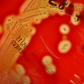
  • Group B Streptococci: Grey-white colonies typically with a narrow zone of β-hemolysis. Bacitracin can help to distinguish it from Group A Strep if needed. Group B are resistant to Bacitracin, while Group A is sensitive, but if being fairly certain from morphology alone, more confirmatory tests can be pursued directly. The CAMP test (Christie–Atkins–Munch-Peterson) is a test that identifies group B β-hemolytic streptococci (Streptococcus agalactiae). The test detects the production of a substance called CAMP factor, which is produced by Group B Streptococcus. The CAMP factor acts synergistically with the beta lysin produced by Staphylococcus aureus to produce a zone of enhanced lysis of sheep or bovine erythrocytes. In the image provided: Streptococcus agalactiae is the vertical line, and S aureus is horizontal line.
  • Group A Streptococci: Smooth or moist greyish-white colonies with wider clear margins of beta-hemolysis. PYR: The Pyrrolidonyl Arylamidase (PYR) test is a rapid biochemical test that detects the ability of bacteria to produce the pyrrolidonyl aminopeptidase enzyme. A color change upon addition of PYR to bacteria is a positive test.
  • Alpha hemolytic Streptococci: Streptococci but a light, greenish agar under the colony on a blood agar plate by partial hemolysis of red blood cells.
  • Enterococcus: Small, smooth, gray or grayish-white, non-hemolytic (γ- hemolytic) colonies.
  • Staphylococci: Spherical gram positive bacteria in clusters. Rapid latex agglutination assay detects PBP2/ (also called PBP2a) in isolates of Staphylococcus. It helps identify methicillin-resistant Staphylococcus aureus (MRSA).
  • Swarming bacteria are generally Proteus. The swarming growth is evidenced as forming a thin filmy layer of concentric circles, which look like the ripples after you throw a rock into a lake
  • Pseudomonas: Pigmented - gray/gray-white with a yellowish tint through green to red or brown.
Date
Source

Own work

Source images used:
Author
Mikael Häggström, M.D. Author info
- Reusing images
- Conflicts of interest:
  None
Mikael Häggström, M.D.

Licensing

w:en:Creative Commons
attribution
This file is licensed under the Creative Commons Attribution 4.0 International license.
You are free:
  • to share – to copy, distribute and transmit the work
  • to remix – to adapt the work
Under the following conditions:
  • attribution – You must give appropriate credit, provide a link to the license, and indicate if changes were made. You may do so in any reasonable manner, but not in any way that suggests the licensor endorses you or your use.

Captions

Microbiology culture algorithm

Items portrayed in this file

depicts

25 July 2023

image/png

File history

Click on a date/time to view the file as it appeared at that time.

Date/TimeThumbnailDimensionsUserComment
current14:58, 27 July 2023Thumbnail for version as of 14:58, 27 July 20235,376 × 4,133 (2.77 MB)Mikael HäggströmCorrection of group B strep
22:40, 26 July 2023Thumbnail for version as of 22:40, 26 July 20235,376 × 4,141 (2.78 MB)Mikael HäggströmUpdate
20:36, 26 July 2023Thumbnail for version as of 20:36, 26 July 20235,376 × 4,181 (2.77 MB)Mikael HäggströmUpdate
22:54, 25 July 2023Thumbnail for version as of 22:54, 25 July 20235,237 × 4,305 (2.75 MB)Mikael HäggströmUploaded a work by {{Mikael Häggström|cat=Medical diagrams}} from {{Own}} with UploadWizard

Global file usage

The following other wikis use this file:

Metadata