Climbing fiber
| Climbing fiber | |
|---|---|
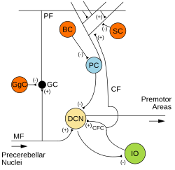
 Microcircuitry of the cerebellum. Excitatory synapses are denoted by (+) and inhibitory synapses by (-). Climbing fiber is shown originating from the inferior olive (green). | |
| Details | |
| Location | Inferior olive and Cerebellum[citation needed] |
| Shape | Unique projection neuron (see text) |
| Function | Unique excitatory function (see text) |
| Neurotransmitter | Glutamate |
| Presynaptic connections | Inferior olive |
| Postsynaptic connections | Purkinje cells |
| Anatomical terms of neuroanatomy | |
Climbing fibers are the name given to a series of neuronal projections from the inferior olivary nucleus located in the medulla oblongata.[1][2]
These axons pass through the pons and enter the cerebellum via the inferior cerebellar peduncle where they form synapses with the deep cerebellar nuclei and Purkinje cells. Each climbing fiber will form synapses with 1-10 Purkinje cells.
Early in development, Purkinje cells are innervated by multiple climbing fibers, but as the cerebellum matures, these inputs gradually become eliminated resulting in a single climbing fiber input per Purkinje cell.
These fibers provide very powerful, excitatory input to the cerebellum which results in the generation of complex spike excitatory postsynaptic potential (EPSP) in Purkinje cells.[1] In this way climbing fibers (CFs) perform a central role in motor behaviors.[3]
The climbing fibers carry information from various sources such as the spinal cord, vestibular system, red nucleus, superior colliculus, reticular formation and sensory and motor cortices. Climbing fiber activation is thought to serve as a motor error signal sent to the cerebellum, and is an important signal for motor timing. In addition to the control and coordination of movements,[4] the climbing fiber afferent system contributes to sensory processing and cognitive tasks likely by encoding the timing of sensory input independently of attention or awareness.[5][6][7]
In the central nervous system, these fibers are able to undergo remarkable regenerative modifications in response to injuries, being able to generate new branches by sprouting to innervate surrounding Purkinje cells if these lose their CF innervation.[8] This kind of injury-induced sprouting has been shown to need the growth associated protein GAP-43.[9][10][11]
See also[edit]
References[edit]
- ^ a b Harting, John K.; Helmrick, Kevin J. (1996–1997). "Cerebellum - Circuitry - Climbing Fibers". Retrieved December 25, 2008.
- ^ Bear, Mark F.; Michael A. Paradiso; Barry W. Connors (2006). Neuroscience: Exploring the Brain (Digitised online by Google Books). Lippincott Williams & Wilkins. p. 773. ISBN 978-0-7817-6003-4. Retrieved December 25, 2008. Image of Parallel fiber
- ^
McKay, Bruce E.; Engbers, Jordan D. T., W. Hamish Mehaffey, Grant R. J. Gordon, Michael L. Molineux, Jaideep S. Bains, and Ray W. Turner; Mehaffey, WH; Gordon, GR; Molineux, ML; Bains, JS; Turner, RW (January 31, 2007). "Climbing Fiber Discharge Regulates Cerebellar Functions by Controlling the Intrinsic Characteristics of Purkinje Cell Output" (PDF). Journal of Neurophysiology. 97 (4): 2590–604. CiteSeerX 10.1.1.325.2405. doi:10.1152/jn.00627.2006. PMID 17267759. Retrieved December 25, 2008.
{{cite journal}}: CS1 maint: multiple names: authors list (link) - ^ "Medical Neurosciences". Archived from the original on January 13, 2012.
- ^ Xu D, Liu T, Ashe J, Bushara KO. Role of the olivo-cerebellar system in timing" J Neurosci 2006; 26: 5990-5.
- ^ Liu T, Xu D, Ashe J, Bushara K. Specificity of inferior olive response to stimulus timing. J Neurophysiol 2008; 100: 1557-61.
- ^ Wu X, Ashe J, Bushara KO. Role of olivocerebellar system in timing without awareness. Proc Natl Acad Sci U S A 2011.
- ^ Carulli D, Buffo A, Strata P (April 2004). "Reparative mechanisms in the cerebellar cortex". Prog Neurobiol. 72 (6): 373–98. doi:10.1016/j.pneurobio.2004.03.007. PMID 15177783. S2CID 18644626.
- ^ Grasselli G, Mandolesi G, Strata P, Cesare P (June 2011). "Impaired Sprouting and Axonal Atrophy in Cerebellar Climbing Fibres following In Vivo Silencing of the Growth-Associated Protein GAP-43". PLOS ONE. 6 (6): e20791. doi:10.1371/journal.pone.0020791. PMC 3112224. PMID 21695168.
- ^ Grasselli G, Strata P (February 2013). "Structural plasticity of climbing fibers and the growth-associated protein GAP-43". Front. Neural Circuits. 7 (25): 25. doi:10.3389/fncir.2013.00025. PMC 3578352. PMID 23441024.
- ^ Mascaro, Allegra; Cesare, P.; Sacconi, L.; Grasselli, G.; Mandolesi, G.; Maco, G.; Knott, G.W.; Huang, L.; De Paola, V.; et al. (2013). "In vivo single branch axotomyinduces GAP-43-dependent sprouting and synaptic remodeling in cerebellarcortex". Proc Natl Acad Sci U S A. 110 (26): 10824–10829. doi:10.1073/pnas.1219256110. PMC 3696745. PMID 23754371.
