
Angular vein
| Angular vein | |
|---|---|
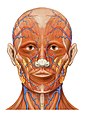
 Veins of the head and neck (angular visible at center right.) | |
 Veins of orbit. | |
| Details | |
| Source | Supraorbital vein |
| Drains to | Facial vein |
| Artery | Angular artery |
| Identifiers | |
| Latin | vena angularis |
| TA98 | A12.3.05.019 |
| TA2 | 4818 |
| FMA | 50893 |
| Anatomical terminology | |
The angular vein is a vein of the face. It is the upper part of the facial vein, above its junction with the superior labial vein. It is formed by the junction of the supratrochlear vein and supraorbital vein, and joins with the superior labial vein. It drains the medial canthus, and parts of the nose and the upper lip. It can be a route of spread of infection from the danger triangle of the face to the cavernous sinus.
Structure[edit]
The angular vein is the upper part of the facial vein, above its junction with the superior labial vein. It anastomoses with the supratrochlear vein,[1] and the supraorbital vein.[2] Its connection with the supraorbital vein forms the superior ophthalmic vein that drains through the orbit.[2] This also connects it with the inferior ophthalmic vein and the cavernous sinus. These do not have valves.[citation needed] The angular vein itself may not contain valves.[3] It receives the lateral nasal veins from the ala of the nose, and the inferior palpebral vein.
The angular vein lies lateral to the angular nerve.[1] It runs obliquely downward by the side of the nose. It passes under zygomaticus major muscle. It joins with the superior labial vein.[citation needed]
Function[edit]
The angular vein drains the medial canthus, and parts of the nose and the upper lip.[4]
Clinical significance[edit]
The angular vein may be affected by a thrombus.[5] This can create problems for endovascular treatment.[5]
Cavernous sinus thrombosis[edit]
Any infection of the mouth or face (such as the danger triangle of the face) can spread to the cavernous sinus via the angular veins. This is particularly as the veins are valveless.[citation needed] This can cause thrombosis. Squeezing pimples in this area should be avoided.[6]
Additional images[edit]
-
 Bloodvessels of the eyelids, front view.
 -
 Lateral head anatomy detail
 -
 Head anatomy anterior view


References[edit]
![]() This article incorporates text in the public domain from page 645 of the 20th edition of Gray's Anatomy (1918)
This article incorporates text in the public domain from page 645 of the 20th edition of Gray's Anatomy (1918)
- ^ a b Caminer, D. M.; Newman, M. I.; Boyd, J. B. (1 April 2006). "Angular nerve: New insights on innervation of the corrugator supercilii and procerus muscles". Journal of Plastic, Reconstructive & Aesthetic Surgery. 59 (4): 366–372. doi:10.1016/j.bjps.2005.09.011. ISSN 1748-6815. PMID 16756251.
- ^ a b Remington, Lee Ann (2012). "11 - Orbital Blood Supply". Clinical Anatomy and Physiology of the Visual System (3rd ed.). Butterworth-Heinemann. pp. 202–217. doi:10.1016/B978-1-4377-1926-0.10011-6. ISBN 978-1-4377-1926-0.
- ^ Zhang, John; Stringer, Mark D. (2010). "Ophthalmic and facial veins are not valveless". Clinical & Experimental Ophthalmology. 38 (5): 502–510. doi:10.1111/j.1442-9071.2010.02325.x. ISSN 1442-9071. PMID 20491800. S2CID 45698367.
- ^ Irmak, M. K.; Korkmaz, A.; Erogul, O. (2004-01-01). "Selective brain cooling seems to be a mechanism leading to human craniofacial diversity observed in different geographical regions". Medical Hypotheses. 63 (6): 974–979. doi:10.1016/j.mehy.2004.05.003. ISSN 0306-9877. PMID 15504564.
- ^ a b Catapano, Joshua S.; Cole, Tyler S.; Albuquerque, Felipe C. (2021). "9 - Hybrid surgical and endovascular treatment". Cerebral Dural Arteriovenous Fistulas. Academic Press. pp. 125–134. doi:10.1016/B978-0-12-819525-3.00004-6. ISBN 978-0-12-819525-3. S2CID 234119518.
- ^ Önerci, T. Metin (2009). Diagnosis in Otorhinolaryngology. Springer-Verlag Berlin Heidelberg. p. 70. ISBN 978-3-642-00498-8.