Fichier:Free-living amebic infections.png
Le contenu de la page n’est pas pris en charge dans d’autres langues.
Une page de Wikipédia, l'encyclopédie libre.

Fichier d’origine(1 365 × 1 170 pixels, taille du fichier : 715 kio, type MIME : image/png)

Ce fichier et sa description proviennent de Wikimedia Commons.

Cette image (de type biologie) devrait être recréée dans un format vectoriel, en tant que fichier SVG. Cela offrirait plusieurs avantages : voir Commons:Media for cleanup pour plus d'informations. Si une version SVG de cette image est déjà disponible, merci de bien vouloir l'envoyer. Après cela, remplacez ce modèle par {{vector version available|nouveau nom d'image.svg}}.

Description

Description
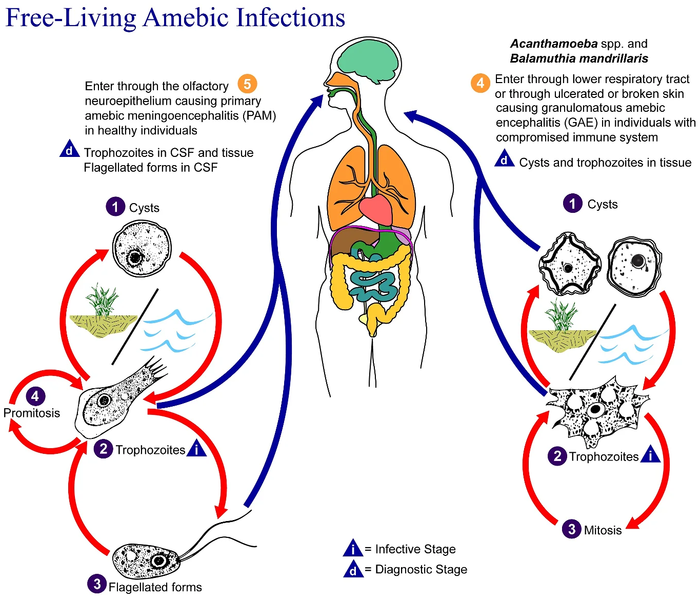
English: This is an illustration of the life cycle of the parasitic agents responsible for causing “free-living” amebic infections.

For a complete description of the life cycle of these parasites, select the link below the image or paste the following address in your address bar: http://www.dpd.cdc.gov/dpdx/HTML/FreeLivingAmebic.htm Free-living amebae belonging to the genera Acanthamoeba, Balamuthia, and Naegleria are important causes of disease in humans and animals. Naegleria fowleri produces an acute, and usually lethal, central nervous system (CNS) disease called primary amebic meningoencephalitis (PAM). N. fowleri has three stages, cysts (1) , trophozoites (2) , and flagellated forms (3) , in its life cycle. The trophozoites replicate by promitosis (nuclear membrane remains intact) (4) . Naegleria fowleri is found in fresh water, soil, thermal discharges of power plants, heated swimming pools, hydrotherapy and medicinal pools, aquariums, and sewage. Trophozoites can turn into temporary flagellated forms which usually revert back to the trophozoite stage. Trophozoites infect humans or animals by entering the olfactory neuroepithelium (5) and reaching the brain. N. fowleri trophozoites are found in cerebrospinal fluid (CSF) and tissue, while flagellated forms are found in CSF.

Acanthamoeba spp. and Balamuthia mandrillaris are opportunistic free-living amebae capable of causing granulomatous amebic encephalitis (GAE) in individuals with compromised immune systems. Acanthamoeba spp. have been found in soil; fresh, brackish, and sea water; sewage; swimming pools; contact lens equipment; medicinal pools; dental treatment units; dialysis machines; heating, ventilating, and air conditioning systems; mammalian cell cultures; vegetables; human nostrils and throats; and human and animal brain, skin, and lung tissues. B. mandrillaris however, has not been isolated from the environment but has been isolated from autopsy specimens of infected humans and animals. Unlike N. fowleri, Acanthamoeba and Balamuthia have only two stages, cysts (1) and trophozoites (2) , in their life cycle. No flagellated stage exists as part of the life cycle. The trophozoites replicate by mitosis (nuclear membrane does not remain intact) (3) . The trophozoites are the infective forms and are believed to gain entry into the body through the lower respiratory tract, ulcerated or broken skin and invade the central nervous system by hematogenous dissemination (4). Acanthamoeba spp. and Balamuthia mandrillaris cysts and trophozoites are found in tissue.
Date
Source

Ce document provient de la Bibliothèque d'images de santé publique (Public Health Image Library) (PHIL) des Centres pour le contrôle et la prévention des maladies , avec le numéro d'identification #3412.

Remarque : toutes les images de la PHIL ne sont pas dans le domaine public ; assurez-vous de vérifier le statut vis-à-vis des droits d'auteur et les crédits aux auteurs et aux fournisseurs de contenus.


العربية | Deutsch | English | македонски | slovenščina | +/−

Auteur
  • Photo Credit:
  • Content Providers(s): CDC/Alexander J. da Silva, PhD/Melanie Moser
Autorisation
(Réutilisation de ce fichier)
English: None - This image is in the public domain and thus free of any copyright restrictions. As a matter of courtesy we request that the content provider be credited and notified in any public or private usage of this image.
Autres versions

Conditions d’utilisation

Public domain
Cette image est l’œuvre des
Centers for Disease Control and Prevention
, division du Département de la Santé et des Services Sociaux des États-Unis, réalisée par un employé dans le cadre de ses activités professionnelles. En tant qu'œuvre du gouvernement fédéral des États-Unis d'Amérique, cette image est placée dans le domaine public.

eesti  Deutsch  čeština  español  português  English  français  Nederlands  polski  slovenščina  suomi  македонски  українська  日本語  中文(简体)‎  中文(繁體)‎  العربية  +/−

Journal des téléversements d’origine

Transféré de en.wikipedia à Commons par Optigan13.

La page de description originale était ici. Tous les noms d'utilisateur qui suivent se rapportent à en.wikipedia.
  • 2006-05-04 01:09 Keenan Pepper 518×435×8 (31658 bytes) Free-living_amebic_infections.gif, converted to [[PNG]] by [[netpbm]].

Légendes

Ajoutez en une ligne la description de ce que représente ce fichier

Éléments décrits dans ce fichier

dépeint

image/png

Historique du fichier

Cliquer sur une date et heure pour voir le fichier tel qu'il était à ce moment-là.

Date et heureVignetteDimensionsUtilisateurCommentaire
actuel2 février 2023 à 11:24Vignette pour la version du 2 février 2023 à 11:241 365 × 1 170 (715 kio)Materialscientisthttps://answersingenesis.org/biology/microbiology/the-genesis-of-brain-eating-amoeba/
20 juillet 2008 à 08:30Vignette pour la version du 20 juillet 2008 à 08:30518 × 435 (31 kio)Optigan13{{Information |Description={{en|This is an illustration of the life cycle of the parasitic agents responsible for causing “free-living” amebic infections. For a complete description of the life cycle of these parasites, select the link below the image

La page suivante utilise ce fichier :

Usage global du fichier

Les autres wikis suivants utilisent ce fichier :

Métadonnées